Konstantinos Theodoridis
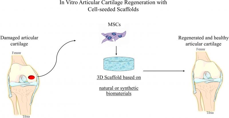
This project aimed to design a 3D-cell Culture Device (3D-CD) for static seeding and cultivation which can be used with several types of scaffolds and to assess and compare static and dynamic seeding and cultivation methods using human adipose tissue. This work was implemented in Medical School of Aristotle University of Thessaloniki.
For more details see Theodoridis, K.; Aggelidou, E.; Manthou, M.-E.; Keklikoglou, K.; Tsimponis, A.; Demiri, E.; Bakopoulou, A.; Mihailidis, A.; Kritis, A. An effective device and method for enhanced cell growth in 3D scaffolds: Investigation of cell seeding and proliferation under static and dynamic conditions. Mater. Sci. Eng. C 2020, 114, 111060, doi:10.1016/j.msec.2020.111060.
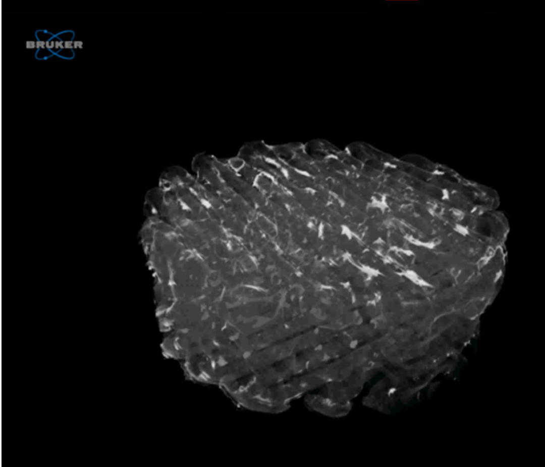
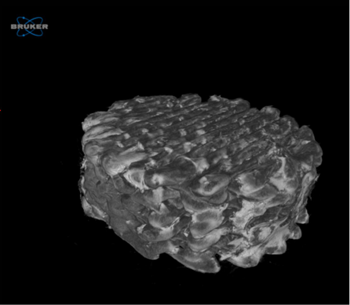
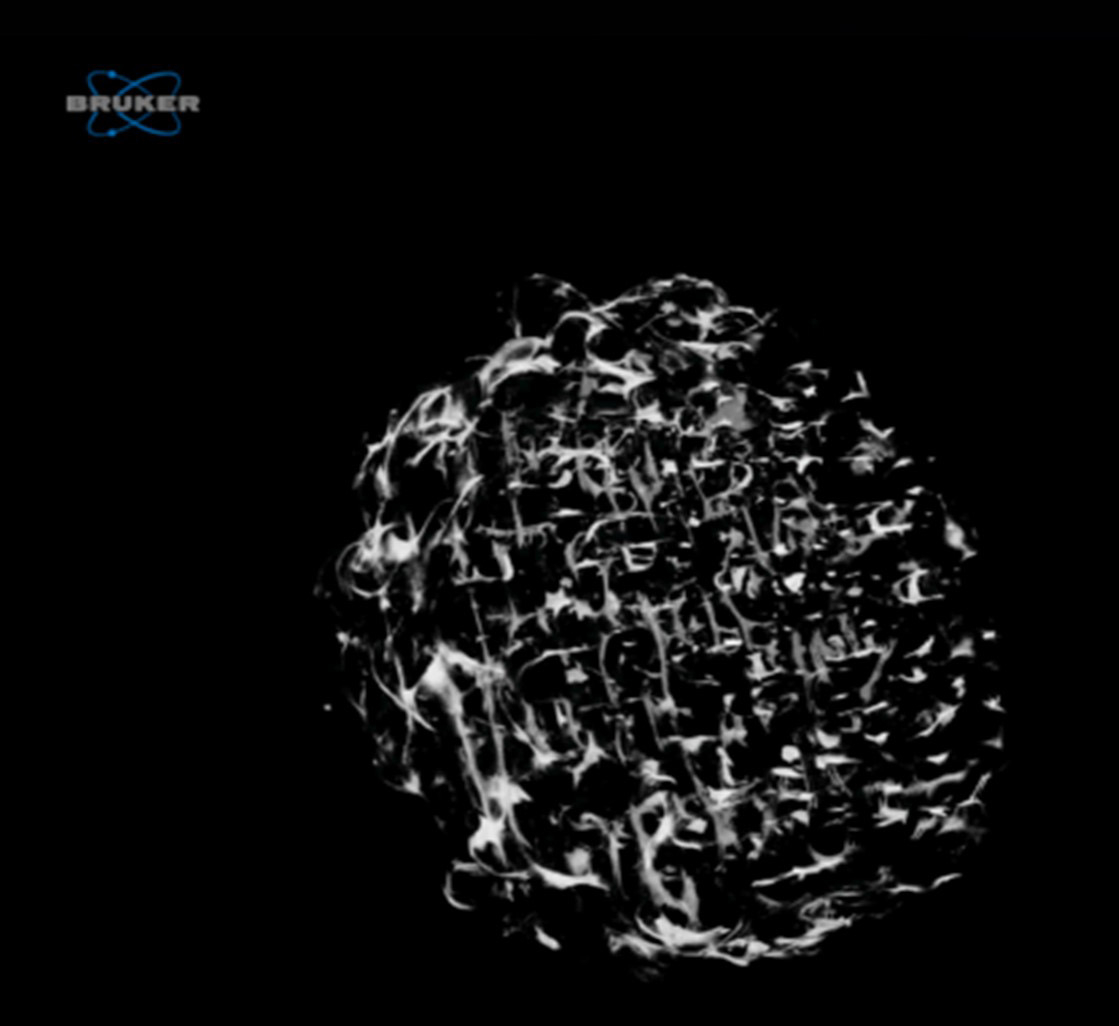
Micro-CT scans were performed to 3D printed scaffolds in order to represent the cell distribution and orientation. 3D rendered images (without the material) of the scanned specimens were created to visually represent the total cartilage tissue formation inside the scaffolds
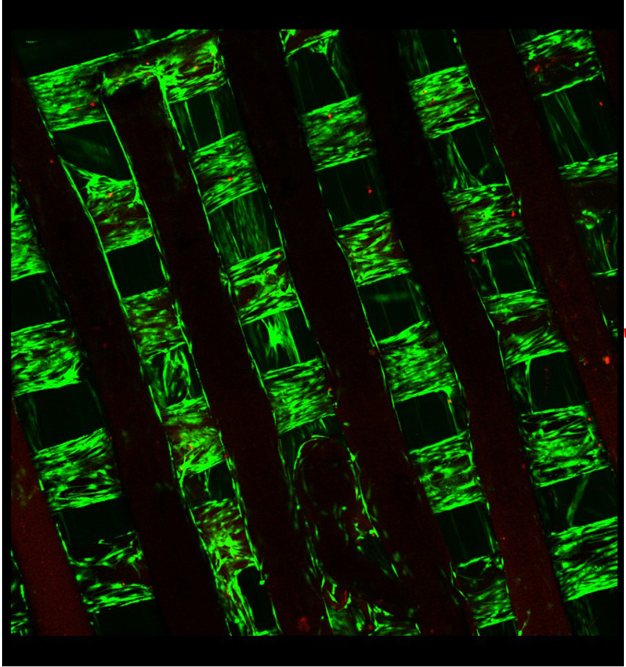
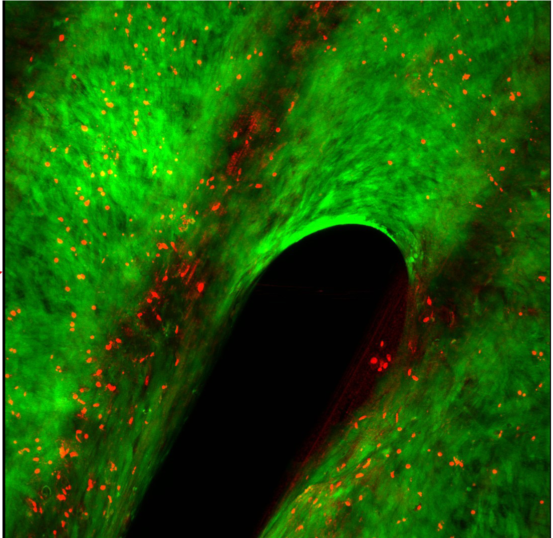
Confocal microscopy was used in order to visualise the adipose derived mesenchymal stem cells seeded in 3D scaffolds and cultured under chondrogenic medium for 21 days. Live (green) / dead (red) fluorescent staining with calcein AM/ethidium homodimer showed high levels of cell viability of cells inside the scaffold's pores.
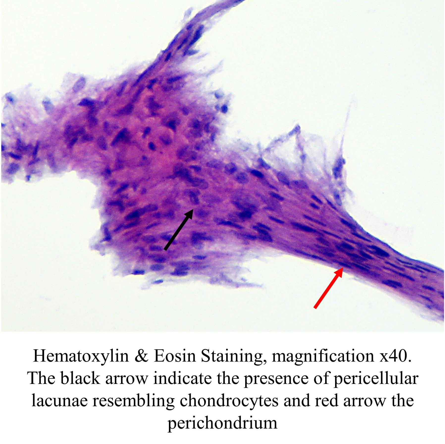
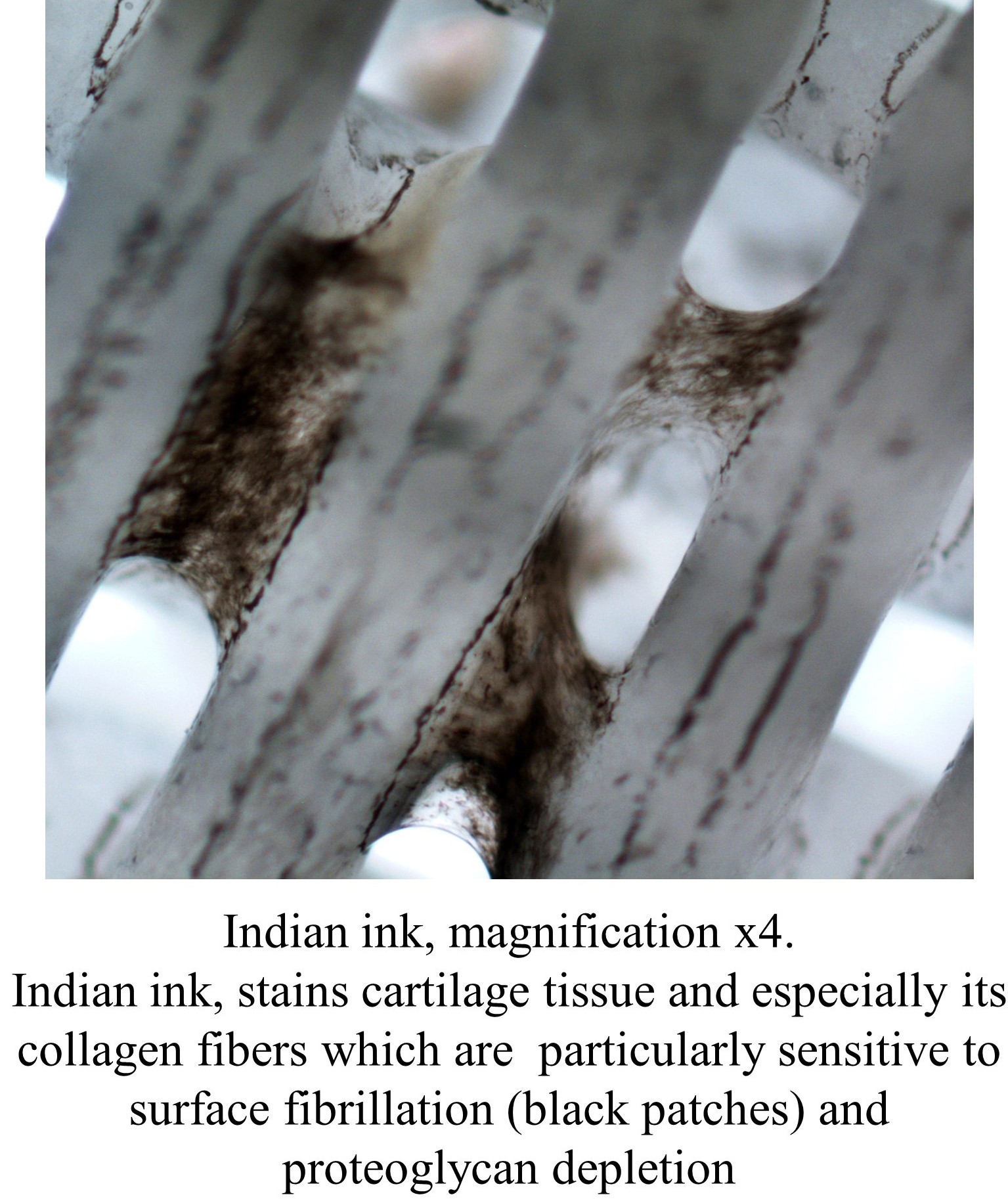
Histological examination was performed to the scaffolds for the cartillage identification.
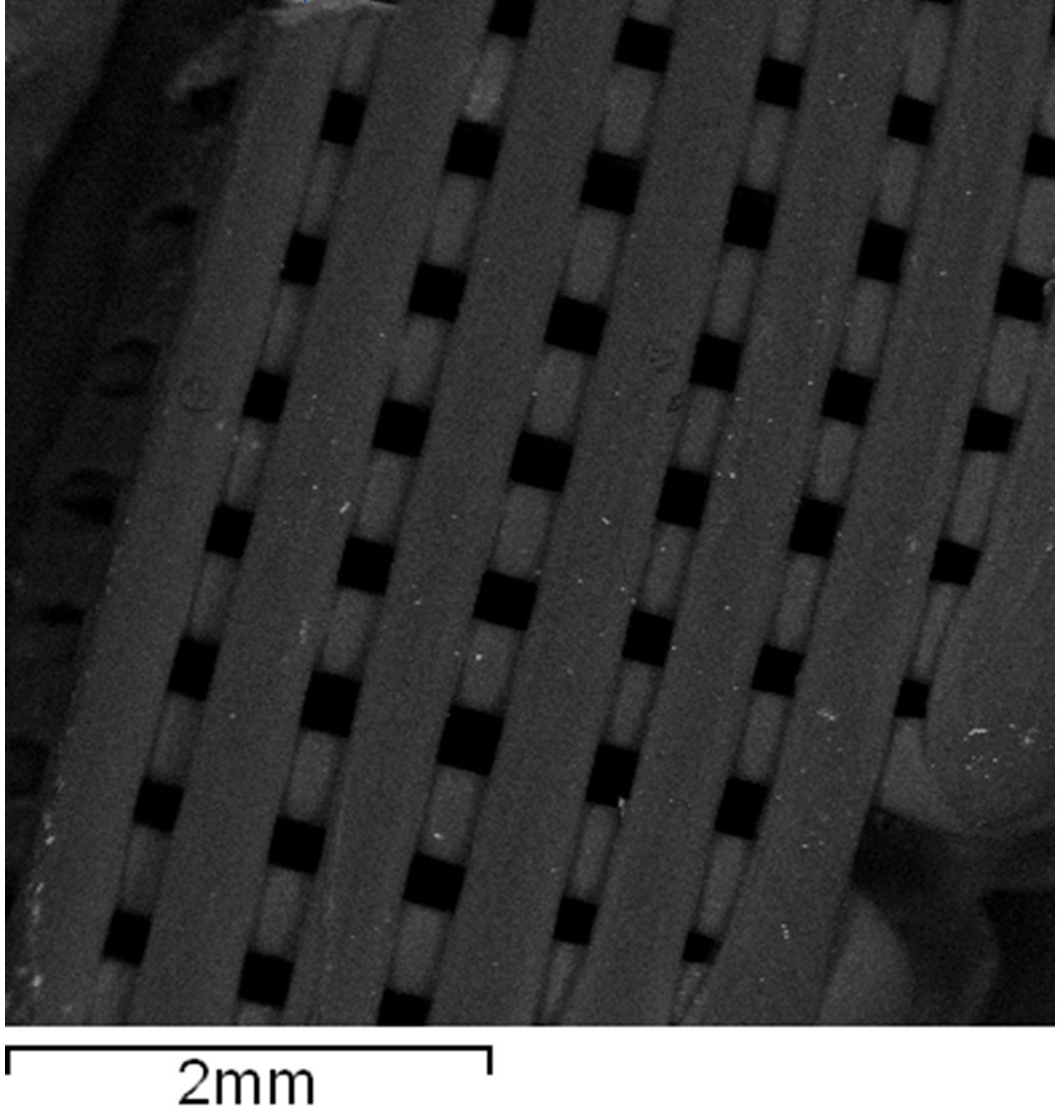
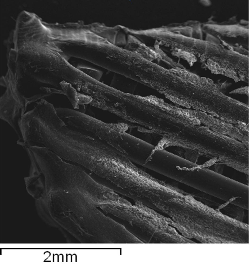
Scanning electron microscopy was performed in order to visualise the null scaffolds as well as the differentiated cells and the extracellular matrix formation