George Mavrothalassitis
Craniosynostosis, the premature ossification of the cranial sutures, is a potentially lethal disease affecting 1 in 2500 newborns. Recent data indicate that ERF haploinsufficiency leads to craniosynostosis, (CRS-4), in both human and mouse. ERF is a transcriptional repressor mamber of the ETS family transcription factors that is inactivated by the FGF/RAS/ERK pathway via phosphorylation and nuclear export. Pharmacological factors that inhibit ERF phosphorylation or nuclear export, increase its suppressor activity. Thus we tested if the remaining Erf activity in the ERF-haploinsufficiency related craniosynostosis mouse model could be augmented by such pharmacological factors to ameliorate the disease phenotype. Newborn animals where treated for 2 months with different dosages and modes of administration. At P65 the animals where sacrificed and scull development, suture closure, calvarium bone thickness and morphology, where analyzed by microcomputed-tomography (microCT).
Details about the imaging techniques used in this project can be found in Keklikoglou, K.; Arvanitidis, C.; Chatzigeorgiou, G.; Chatzinikolaou, E.; Karagiannidis, E.; Koletsa, T.; Magoulas, A.; Makris, K.; Mavrothalassitis, G.; Papanagnou, E.-D.; Papazoglou, A.S.; Pavloudi, C.; Trougakos, I.P.; Vasileiadou, K.; Vogiatzi, A. Micro-CT for Biological and Biomedical Studies: A Comparison of Imaging Techniques. J. Imaging 2021, 7, 172. https://doi.org/10.3390/jimaging7090172
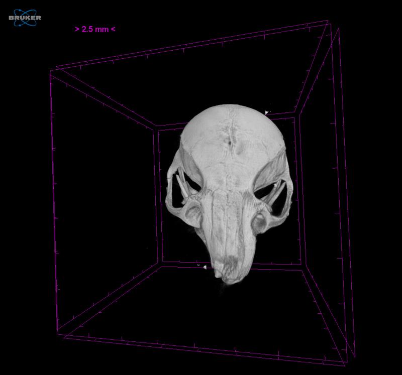
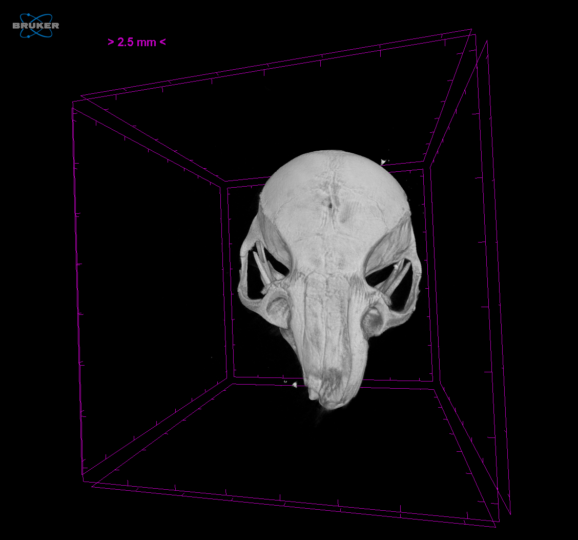
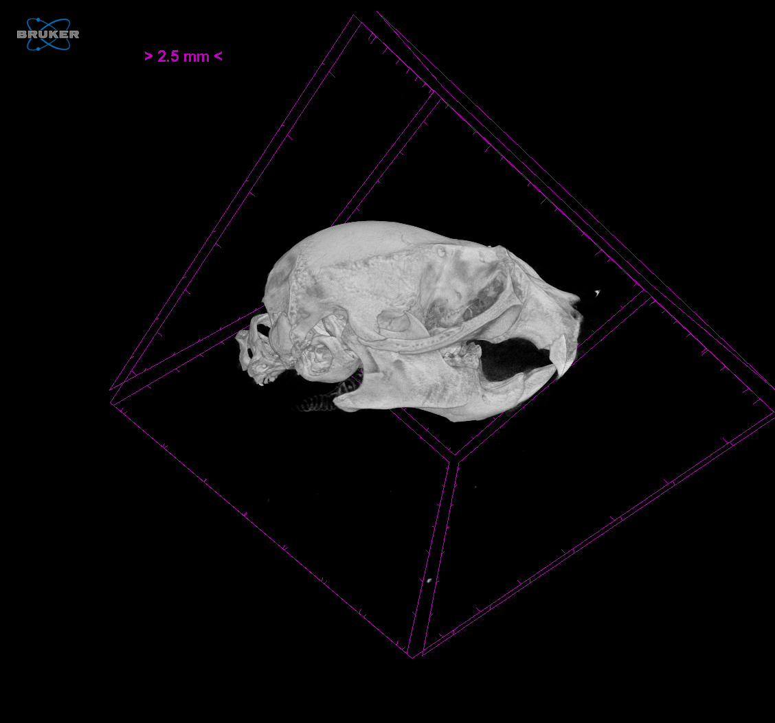
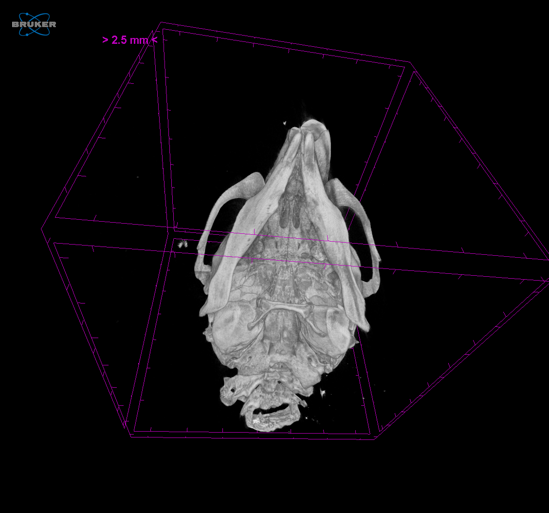
Micro-CT was performed to the skull of a mouse (Mus musculus). This specimen was preserved in 100% ethanol. For this specimen, no contrast agent was used. Scan was performed with a Skyscan 1172 at a voltage of 75kV and 131μΑ with an an aluminium filter of 0.5mm for a half rotation of 180o . This specimen was scanned at a pixel size of 13.79μm with an exposure time of 1435ms. This scan was performed to a ErfloxP/- craniosynostosis animal. Specimen was provided by George Mavrothalassitis (FORTH) and scanned by Niki Keklikoglou.
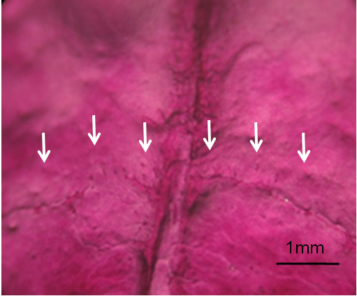
Coronal Sutures of P65 mouse calvaria stained with Alizarin Red and Alcian Blue. The white arrows indicate the position of the ossified suture in the ErfloxP/−(CRS‐4) craniosynostosis animals. The sagittal (vertical) suture is visible.
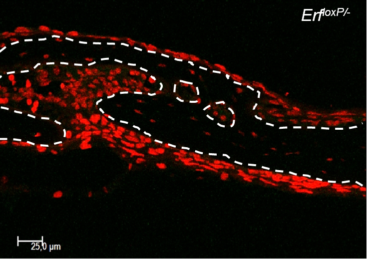
Confocal microscopy of coronal sutures transverse cryosection from P15 mouse calvaria stained with BrdU for cellular to evaluate proliferation and TOPRO-3 to identify the nuclei. Doted lines indicate the position of the parietal and frontal bones.