Efstratios Karagiannidis
Micro‐CT and histological imaging were utilised to evaluate the impact of aspirated thrombus burden on the post‐aspiration clinical and angiographical outcomes of patients with ST‐Elevated Myocardial Infarction (STEMI).
Details about the imaging techniques used in this project can be found in Keklikoglou, K.; Arvanitidis, C.; Chatzigeorgiou, G.; Chatzinikolaou, E.; Karagiannidis, E.; Koletsa, T.; Magoulas, A.; Makris, K.; Mavrothalassitis, G.; Papanagnou, E.-D.; Papazoglou, A.S.; Pavloudi, C.; Trougakos, I.P.; Vasileiadou, K.; Vogiatzi, A. Micro-CT for Biological and Biomedical Studies: A Comparison of Imaging Techniques. J. Imaging 2021, 7, 172. https://doi.org/10.3390/jimaging7090172
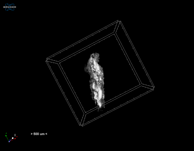
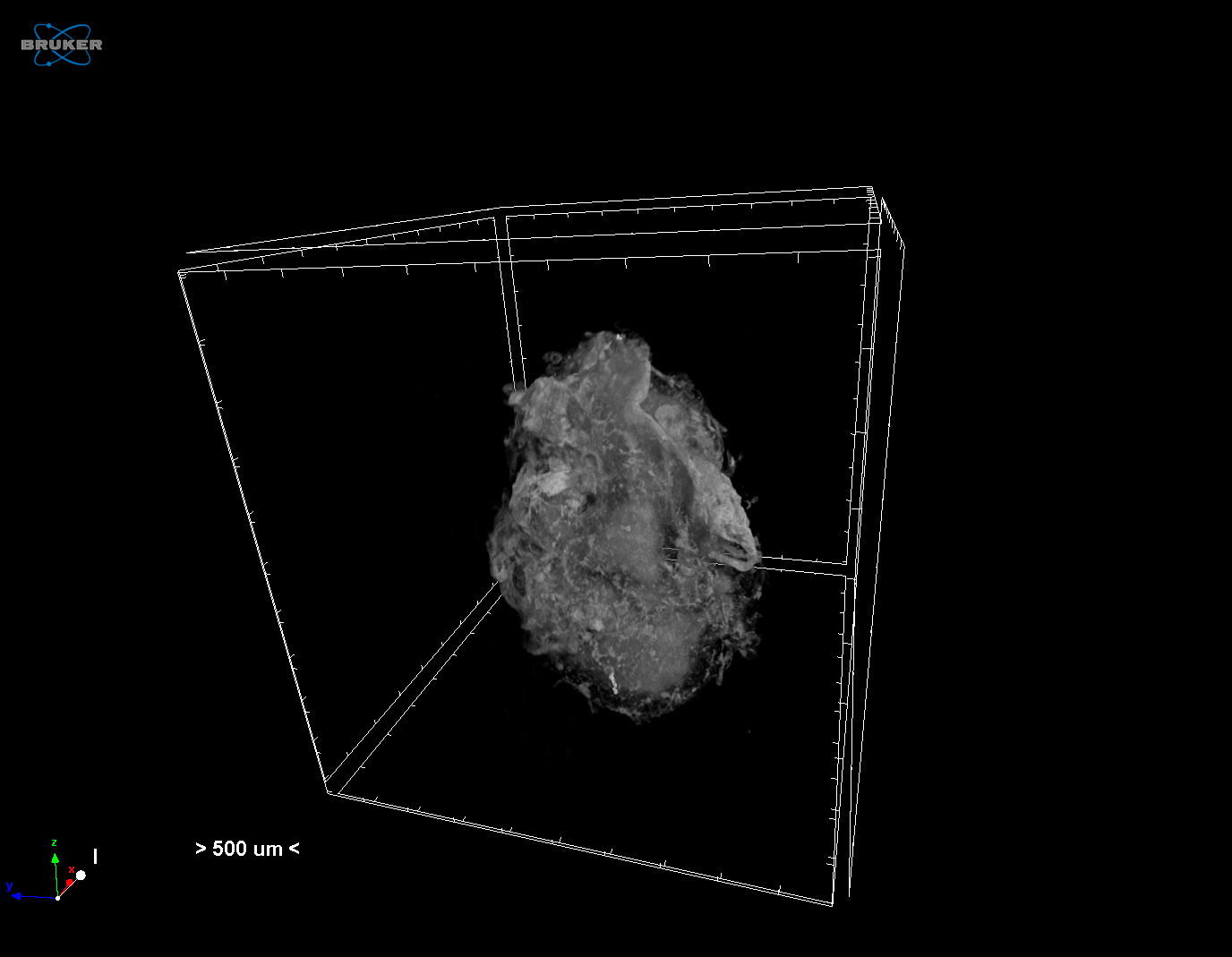
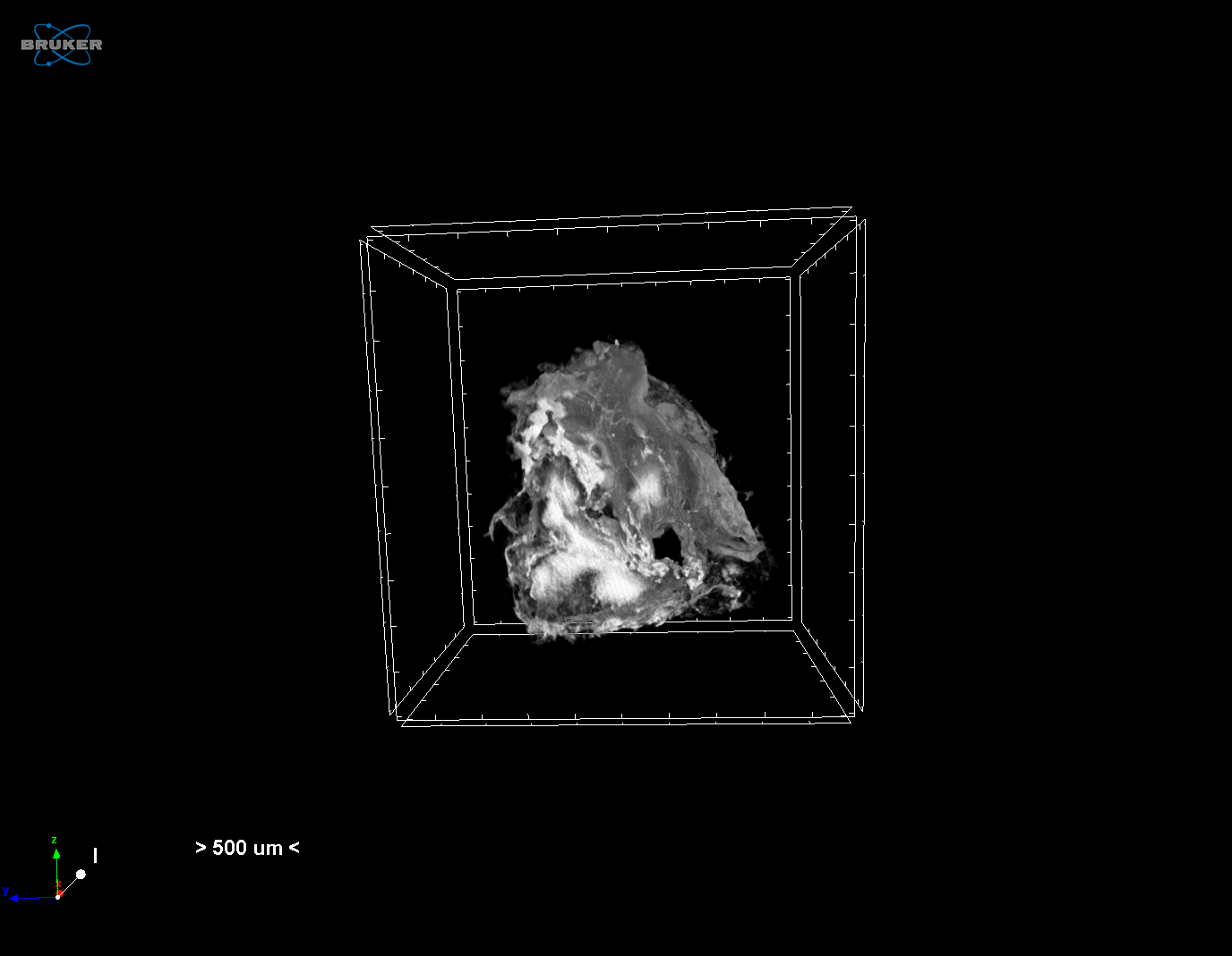
Micro‐CT was performed for the evaluation of the impact of aspirated thrombus burden on the post‐aspiration clinical and angiographical outcomes of patients with ST‐Elevated Myocardial Infarction (STEMI). Thrombotic materials were fixed in formalin and preserved in ethanol. Specimens were stained with 0.3% PTA dissolvded in 70% ethanol. Scans were performed with a Skyscan 1172 at a voltage of 50kV and 198μΑ without filter for a full rotation of 360o . Specimen was provided by Efstratios Karagiannidis (AUTH) and scanned by Eva Chatzinikolaou.

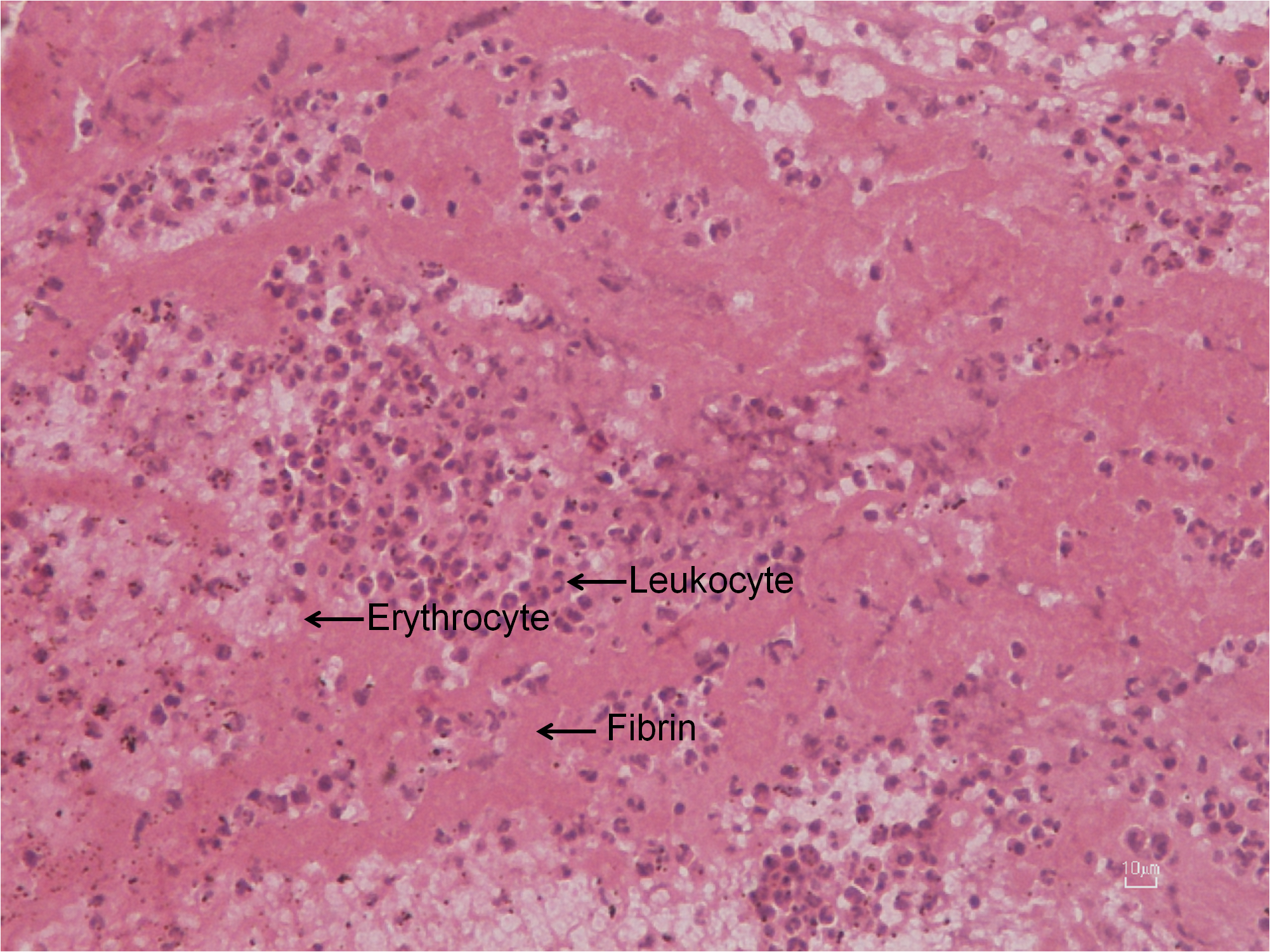
Histological examination was performed to complement the micro‐CT analysis, and it revealed a thrombus characterised by fibrin/erythrocytes (occupied 70% of the total area of the thrombus) and leukocytes (30%) after staining the formalin‐fixed paraffin‐embedded specimen with hematoxylin and eosin.